Imaging technology
Micro CT
Nano CT
Scanning Electron Microscopy (SEM)
Transmission Electron Microscopy (TEM)
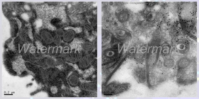
Immunoelectron Microscopy (IEM)
Cryo-Electron Microscopy (Cryo-EM)
Liquid-Phase Electron Microscopy (Liquid EM)
In Vivo Fluorescence Imaging
Immunoelectron microscopy is a highly precise and sensitive technique that combines the specificity of antigen-antibody reactions with the high resolution of electron microscopy to perform localization analysis of antigens at the subcellular and ultrastructural levels.

Hong Kong Office:224 Waterloo Road, Kowloon Tong, Hong Kong (inside the Baptist University campus)
Shanghai Office:2F, 1788 Caoyang Road, Putuo District, Shanghai
Shanghai Office:2F, 1788 Caoyang Road, Putuo District, Shanghai
copyright © ROYAL BIOTECH All rights reserved.
